Multiple sclerosis is a chronic disease that attacks the central nervous system. It is thought to be an autoimmune disease in which the body’s own defence system attacks the myelin that surrounds nerve fibers in the brain.
The course of disease that a particular patient will experience is hard to predict. We are interested in utilizing advanced MR imaging to characterize white matter injury, and to understand the relationship between the position of lesions and the deficits created by those lesions.
Disruption of White Matter Circuits in Multiple Sclerosis
 |
|---|
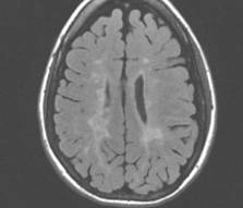
| White matter lesions in FLAIR MRI |
Focal and diffusion white matter lesions are common in multiple sclerosis, but more study is needed to understand the importance of the distribution of such changes across the brain. Focal white matter lesions are common across normal aging as well as a range of diseases and syndromes, and are associated with decreased motor skills and decreased cognitive performance. Conventional magnetic resonance imaging (MRI) is now a standard clinically accepted mechanism to assess and to monitor white matter (WM) lesion presence and activity in the brain. Currently studies to assess clinical disability are predominantly based upon lesion burden, that is, the total number or total size of plaques in the entire brain, or change in lesion burden as indicated by new lesions.
Our research aims to take advantage of recent advances in magnetic resonance imaging and post-acquisition image analysis in order to improve our understanding of the role of the underlying white matter connectivity in assessing the impact of WM abnormalities.
Recent advances in diffusion tensor magnetic resonance imaging (DT-MRI) have provided the image acquisition technology to enable the new in vivo assessment of the role of white matter architecture of the human brain, which we propose here.
The primary objective of this project is to create and utilize new image analysis techniques to enable the identification of white matter fiber tract segments that are disrupted by focal white matter lesions, and to identify the essential patterns of WM disruption that are most highly correlated with decreased cognitive performance. These results will significantly improve our understanding of the relation between the anatomical location of focal white matter abnormalities, the connectivity of the white matter and cognitive impairment.
Segmentation of Multiple Sclerosis Lesions from MRI
We have developed effective segmentation algorithms for multiple sclerosis lesions by utilizing a combination of MRI signal intensity and spatial context from a digital anatomical atlas (Warfield et al. 1995; Warfield et al. 2000). This combination of segmentation and nonrigid registration has proved to be a powerful paradigm for image analysis.
Tractography in the presence of lesions
 |
|---|
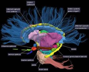
| High resolution single subject atlas of white matter fiber tracts |
In order to identify the fiber tracts disrupted by particular lesions, we have developed a tract clustering approach (Maddah et al. 2005; Maddah, Wells, Warfield et al. 2007; Maddah, Wells, Westin et al. 2007; Maddah et al. 2008). This algorithm identifies fiber tracts in a particular subject, and can utilize a model of normal fiber tracts such as are encoded in our single subject atlas illustrated below:
White matter lesions disrupt the structure of the white matter. This may make it challenging to detect fiber tracts, but sophisticated tractography algorithms are able to identify fiber tracts despite regions with reduced signal due to the presence of lesions (Goldberg-Zimring et al. 2005).
References
- O Commowick, S K Warfield. A continuous STAPLE for scalar, vector, and tensor images: an application to DTI analysis. IEEE Trans Med Imaging, 28(6):838-46, 2009.
- M Maddah, W E Grimson, S K Warfield, and W M Wells. A unified framework for clustering and quantitative analysis of white matter fiber tracts. Med Image Anal., 12(2):191-202, April 2008.
- O Commowick, P Fillard, O Clatz, S K Warfield. Detection of DTI white matter abnormalities in multiple sclerosis patients. Med Image Comput Comput Assist Interv Int Conf Med Image Comput Comput Assist Interv., 11(Pt 1):975-82, 2008.
- M Maddah, W M Wells, S K Warfield, C F Westin, and W E Grimson. Probabilistic clustering and quantitative analysis of white matter fiber tracts. Inf Process Med Imaging, 20:372-83, 2007.
- M Maddah, W M Wells, C F Westin, W E Grimson, and S K Warfield. A Spatial Model of White Matter Fiber Tracts. Paper read at ISMRM, at Berlin 2007.
- D Goldberg-Zimring, S K Warfield. Novel image processing techniques to better understand white matter disruption in multiple sclerosis. Autoimmun Rev., 5(8):544-8, 2006.
- Y Wu, S K Warfield, I L Tan, W M Wells 3rd, D S Meier, R A van Schijndel, F Barkhof, C R Guttmann. Automated segmentation of multiple sclerosis lesion subtypes with multichannel MRI. Neuroimage, 32(3):1205-15, 2006.
- D Goldberg-Zimring, A U Mewes, M Maddah, and S K Warfield. Diffusion tensor magnetic resonance imaging in multiple sclerosis. J Neuroimaging, 15 (4 Suppl):68S-81S, 2005.
- M Maddah, A U Mewes, S Haker, W E Grimson, and S K Warfield. Automated atlas-based clustering of white matter fiber tracts from DTMRI. Med Image Comput Comput Assist Interv Int Conf Med Image Comput Comput Assist Interv., 8(Pt 1):188-95, 2005.
- X Wei, C R Guttmann, S K Warfield, M Eliasziw, J R Mitchell. Has your patient’s multiple sclerosis lesion burden or brain atrophy actually changed? Mult Scler., 10(4):402-6, 2004.
- D Goldberg-Zimring, A Achiron, S K Warfield, C R Guttmann, H Azhari. Application of spherical harmonics derived space rotation invariant indices to the analysis of multiple sclerosis lesions’ geometry by MRI. Magn Reson Imaging, 22(6):815-25, 2004.
- X Wei, S K Warfield, K H Zou, Y Wu, X Li, A Guimond, J P Mugler 3rd, R R Benson, L Wolfson, H L Weiner, C R Guttmann. Quantitative analysis of MRI signal abnormalities of brain white matter with high reproducibility and accuracy. J Magn Reson Imaging, 15(2):203-9, 2002.
- R A Sperling, C R Guttmann, M J Hohol, S K Warfield, M Jakab, M Parente, E L Diamond, K R Daffner, M J Olek, E J Orav, R Kikinis, F A Jolesz, H L Weiner. Regional magnetic resonance imaging lesion burden and cognitive function in multiple sclerosis: a longitudinal study. Arch Neurol., 58(1):115-21, 2001.
- S K Warfield, M Kaus, F A Jolesz, and R Kikinis. Adaptive, template moderated, spatially varying statistical classification. Med Image Anal., 4 (1):43-55, 2000.
- C R Guttmann, R Kikinis, M C Anderson, M Jakab, S K Warfield, R J Killiany, H L Weiner, F A Jolesz. Quantitative follow-up of patients with multiple sclerosis using MRI: reproducibility. J Magn Reson Imaging, 9(4):509-18, 1999.
- S K Warfield, J Dengler, J Zaers, C R Guttmann, W M Wells, G J Ettinger, J Hiller, and R Kikinis. Automatic identification of gray matter structures from MRI to improve the segmentation of white matter lesions. J Image Guid Surg., 1(6):326-38, 1995.